
Priyadharshini Hills P. O
Athirampuzha
Kottayam, Kerala, India-686560
P +91 2732120, +91 9447 391 168
E cta@mgu.ac.in
F +91 481 271 009/02

on October 3rd to 6th2013
on September 6th to 9th 2013
on October 5th to 8th 2012
The origin of mass spectrometry is from the discovery of positive rays in gas discharge tube experiments to study the conduction of electricity by gases at very low pressure. Eugen Goldstein in his experiments on discharge tube using perforated cathode found that there are a beam of positively charged particles originating from the anode, moving towards the cathode. He called these as “Kanalstrahlen” meaning Canal rays. Wilhem Wien found that these rays can be deflected by electric and magnetic fields according to their mass to charge (m/z) ratio. The m/z ratio of the particles in the canal rays were found to depend on the nature of the gas taken in the discharge tube.
J.J. Thomson carried out further experiments on these positive rays to find out the particle responsible for the positive charge. He found that each element possesses its own characteristic positive ion and some possess more than one. This led to the discovery of isotopes. Thomson discovered two isotopes of neon, 20Ne and 22Ne. He isolated these isotopes using his positive ray parabolae apparatus, the first mass spectrometer in history. He applied electric and magnetic fields to the positive rays generated in the discharge tube in a perpendicular fashion, which separated the ions according to the m/z ratio.
Later on Francis William Aston improved the positive ray parabolae apparatus and built his mass spectrograph, the ancestor of modern spectrometers. He subjected the ions to electric and magnetic fields separately making the ions to deflect and focus on a particular point on the photographic plate. Thus he could scan a particular mass range by adjusting the fields.
Earlier the mass spectrometers were predominantly used by inorganic chemists to discover and characterize different isotopes of elements. It was observed that there were background spectra which were characteristic of the lubricants and other organic fluids associated with the instrument. There begins organic mass spectrometry.
Now there has been an explosion of knowledge in the field of mass analysis. Mass spectrographs became mass spectrometers. Now it can be used to get structural information of a wide range of molecules including biomolecules which is made possible by the development of different ionization techniques. The first application of mass spectrometry to amino acids and polypeptides was reported in 1958 by Carl-Ove Andersson.
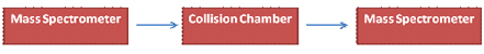
Nowadays mass spectrometers are used as detecting systems in chromatographic techniques in the so called hyphenated techniques- GC-MS, LC-MS etc. Being the combination of two micro analytical techniques, they are superior to the conventional mass spectrometry. By the advent of tandem mass spectrometry, it is now possible to take the mass spectrum of a mass spectrum.
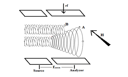
2. Ionization and accelerating chamber
3. Mass analyzer
4. Ion collector and amplifier
5. Recorder
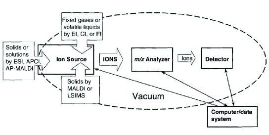
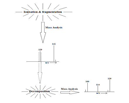
| | In a mass spectrometer, some form of energy is transferred to the molecule and they are ionized and fragmented. |
| | The ions formed are accelerated by an electric field and separated according to their m/z ratio. |
| | These sorted ions are led to a detector which can count the number of ions striking it and it produces a current proportional to it. |
| | The detector output is amplified and passed to the recorder |
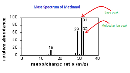
| | The mass spectrum is obtained from the recorder which is a plot of percentage of abundance of the ions Vs m/z ratio, the most abundant ion being given a value for percentage of abundance 100. |
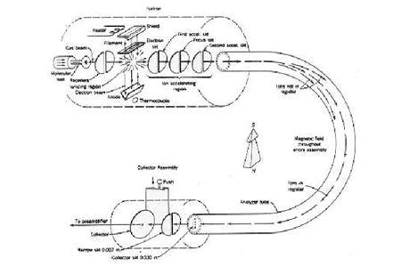
 Schematic Diagram of the processes in a mass spectrometer (ref: Introduction to Mass spectrometry, J. Throck Watson, 3rd edition)
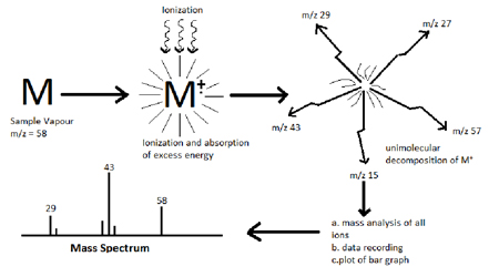
M (g) +e ![]() M+.+2e
M+.+2e
m1+ ![]() m2+ + neutral molecule
m2+ + neutral molecule
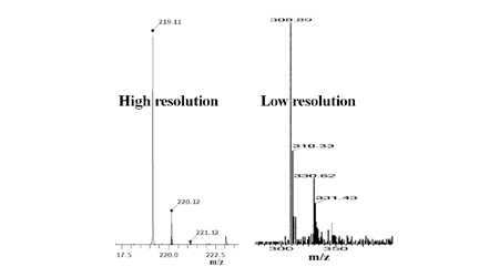
Resolution = M/M
M and M are the m/z values of two closely lying peaks in the spectrum
Ionization method |
Typical |
Sample introduction |
Mass range |
Method highlights |
Electron impact (EI) |
Relatively |
GC or liquid/solid probe |
To 1000 Da |
Hard method, versatile, provide structural info |
Chemical ionization (CI) |
Relatively |
GC or liquid/solid probe |
To |
Soft method, molecular ion peak [M+H]+ |
Electrospray ionization (ESI) |
Peptides, proteins, non volatile molecules |
Liquid chromatography or syringe |
To 200000 Da |
Soft method, ions often multiply charged |
Fast atom bombardment (FAB) |
Carbohydrates, peptides, organome- |
Sample mixed |
To 6000 Da |
Soft method, harder than ESI and MALDI |
Matrix Assisted Laser Desorption Ionization (MALDI) |
Peptides, proteins, nucleotides |
Sample mixed |
To 500000 Da |
Soft method, very high mass |
- Magnetic field only
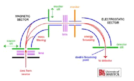
- Double Focusing (Electric and Magnetic fields)
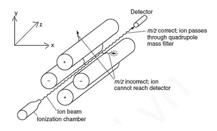
- Quadrupole Mass Filter
- Quadrupole Ion Storage (Ion Trap)

Quadrupole |
Unit mass resolution, fast scan, low cost |
Sector (magnetic sector and double focusing) |
High resolution, exact mass |
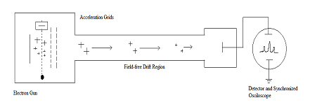
Time-Of-Flight (TOF) |
Theoretically no limitation for m/z maximum, high throughput |
Ion Cyclotron Resonance |
Very high resolution, exact mass, perform ion chemistry |
2. Introduction to Mass spectrometry, J. Throck Watson, 3rd edition.
3. Spectroscopy, Robert M. Silverstein, Francis X. Webster

Group | Collabrotions | Photos | Contact
| CTAMGU © 2012 | Design & Developed By : ITS Developers |